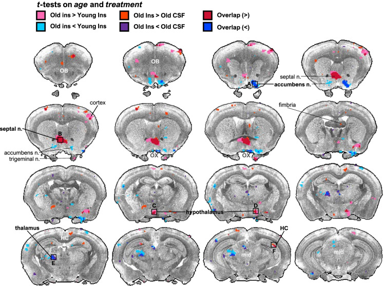
Fig. 4.
Central insulin elicits distinct brain regional effects on the rs-fMRI signal in old, but not young rats. Young (n = 8) and old rats (n = 9) were assigned to a crossover design whereby animals were randomly assigned to either ICV CSF or insulin first and then received the other treatment 1 week later. The rs-fMRI imaging session was 1 h in length and BOLD signal intensity was measured in extracted ROIs. No differences were observed in ΔBOLD signal between young and old rats treated with CSF. However, ICV insulin surprisingly failed to significantly alter the BOLD signal in any region in young rats but led to several dynamic changes in BOLD signal across the brain in old. Specifically, ICV-insulin treated animals exhibited significantly increased mean BOLD signal intensity in septal nucleus, hypothalamus, and hippocampus and decreased intensity in thalamus and nucleus accumbens and compared to old CSF and young insulin treated animals. Highlighted regions indicat significantly different BOLD signal intensity between age (independent t-test on ΔBOLD signal, treatment (paired t-test within age group), or both determined; where pink, aged insulin > young insulin; orange, aged insulin > aged aCSF; red, aged insulin > young insulin and aged CSF; turquoise, aged insulin < young insulin; indigo, aged insulin < aged aCSF; blue, aged insulin < young insulin and aged aCSF. Regions of interest (ROIs) were identified and projected in Waxholm space

