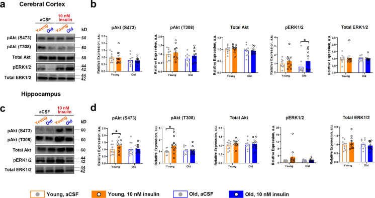
Fig. 5.
Ex vivo insulin exposure in rat cerebral cortex and hippocampus and modulation of PI3K/Akt and MAPK signaling pathways. Approximately 20 mg of cerebral cortex and hippocampus was each collected from young and old rats immediately after sacrifice and incubated for 2 min either in aCSF or 10 nM insulin in a 37 °C incubator, 5% CO2 (n = 10 per group) following 10-min untreated thermal and biological equilibration (12-min total incubation time). a–b Tissue responsiveness to 10 nM insulin in cortex, indicated by phosphorylation of Akt on Ser473 or Thr308 residues indicated no effect in altering either pAkt S473 or T308 phosphorylation. However, insulin tended to increase pErk levels in cortex, but this was only significant in old animals. c–d In hippocampus, insulin was able to significantly activate pAkt in young, as evidenced by increased phosphorylation at S473 and T308, but not in old. There were also no significant effects detected in the Erk pathway, collectively suggesting discordance regarding insulin effects with age among in vivo and ex vivo assays. Data were analyzed as a paired t-test within each age group. Representative Western blots are shown. Bar graphs represent mean ± SE. *p ≤ 0.05

