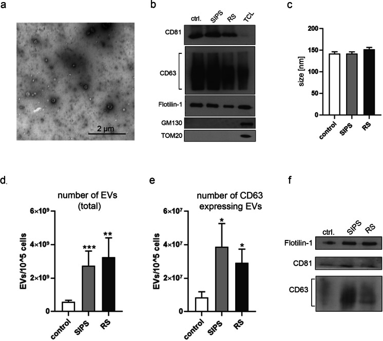
Fig. 1.
Characterization of extracellular vesicles secreted by VSMCs. a Isolated EVs, obtained after ultracentrifugation of conditioned medium, observed in TEM; b Western blot analysis of common exosome markers (CD81, CD63, and Flotilin-1) in EV fraction (20µg of each sample). The purity of isolated EVs was confirmed by lack of expression of Golgi (GM130) and mitochondrial (TOM20) markers, TLC — total cell lysate; c Size measurement of EVs using NanoSight nanoparticle tracking analysis (NTA). d The total number of EVs secreted by young and senescent cells measured by NTA; EVs from at least 9 independent isolations from each experimental conditions, control, SIPS and RS, cells were analyzed. e Comparison of the number of CD63 positive EVs secreted by VSMCs (ExoElisa); EVs from at least 9 independent isolations were analyzed. f Representative blots presenting the differences in the level of CD81, CD63, and Flotilin-1 protein in EVs secreted by 2 × 10⁵ young and senescent cells

