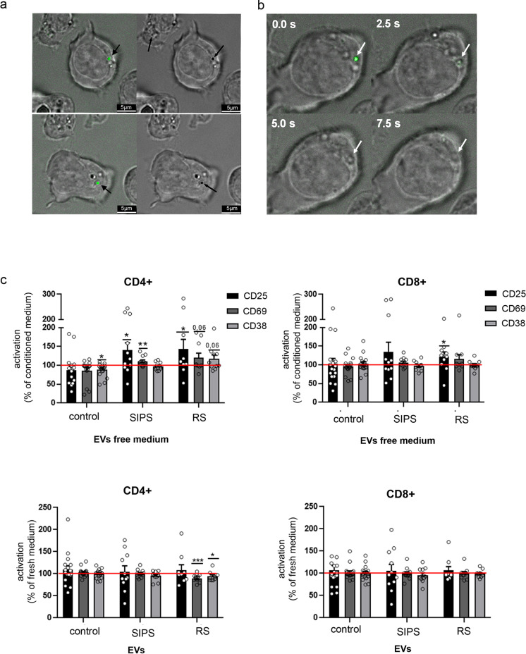
Fig. 5.
The influence of VSMCs-EVs on T cell activation. a Visualization of the uptake of VSMC-EVs by CD3 + cells. T cells were cocultured with CFSE stained EVs for 4 h in 37 °C and analyzed in bright-field merged with fluorescence microscopy. b Time laps analysis of CFSE stained EVs incorporated by T cells; c Expression of CD25, CD69, and CD38 in CD4 + and CD8 + subsets of T cells activated in the presence of EV-free medium or medium supplemented with EVs collected from control and senescent VSMCs. The number of cells expressing CD25, CD69, or CD38 was measured after 24 h of activation and normalized to the number of activated T cells in full conditioned medium (medium containing EVs). T cells isolated from at least 9 donors were analyzed; for statistical analysis t-test was used

