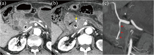
FIGURE 2.

(a) Contrast‐enhanced computed tomography reveals narrowing in the gastroduodenal artery near the duckbill‐shaped anti‐reflux metal stent. (b) Contrast‐enhanced computed tomography reveals dilatation in the peripheral gastroduodenal artery compared to (a), considered to represent a pseudoaneurysm (yellow arrow). (c) Narrowing and irregular dilatation (red arrow) are found in the gastroduodenal artery and determined to represent pseudoaneurysm.
