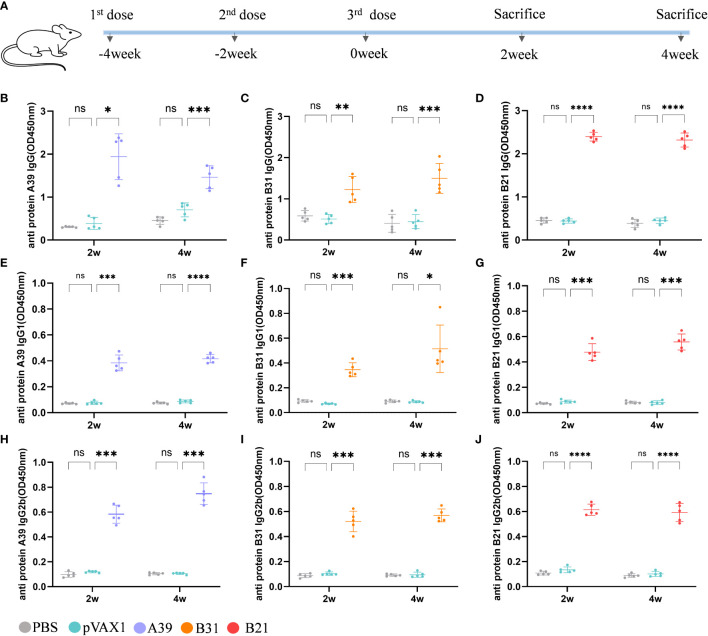
Figure 2.
Analysis of antibodies in the serum of immunized mice. Levels of antibody detected by ELISA at 2 and 4 weeks after immunization. Samples were diluted 1/1000 in PBS buffer. (A) Schedule for DNA vaccine immunization in mice. (B-D) Anti A39, B31, B21 protein IgG antibody; (E-G) Anti A39, B31, B21 protein IgG1 antibody; (H-J) Anti A39, B31, B21 protein IgG2b antibody. Data are shown as mean±SD, n = 5. Statistical analyses performed by two-way ANOVA (ns, No significance; *P < 0.05; **P < 0.01; ***P < 0.001; ****P < 0.0001).

