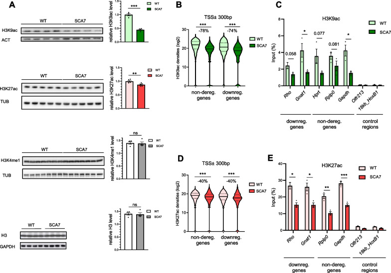
Fig. 2.
Genome-wide analysis H3K9 and H3K27 acetylation at gene promoters in SCA7 retina. A Western blot analyses showing the significant decrease of H3K9ac and H3K27ac levels in SCA7 retina compared to WT, while H3K4me1 and unmodified H3 levels are not affected. Data are normalized to the level of control proteins (actin (ACT), tubulin (TUB) and GAPDH), expressed as mean ± SEM (n = 4–7 mice/genotype) and analyzed using two-tailed Student’s t-test. B Violin plots showing the reduction of H3K9ac densities on regions ± 300 bp around the transcription start site (TSS ± 300 bp) of non-deregulated and downregulated genes in SCA7 retina, compared to WT. C ChIP-qPCR analysis showing the decrease of H3K9ac occupancy (% of the input) on the TSS region of representative downregulated and non-deregulated genes in SCA7 retina compared to WT. Silent regions are used as controls. D Violin plots showing the reduction of H3K27ac densities on the TSS ± 300 bp regions of non-deregulated and downregulated genes in SCA7 retina, compared to WT. E ChIP-qPCR analysis showing the decrease of H3K27ac occupancy on the TSS region of representative downregulated and non-deregulated genes in SCA7 retina compared to WT. Silent regions are used as controls. Data in B and D were analyzed using Mann–Whitney test. Data in C and E are normalized as percentage of input DNA signal, expressed as mean ± SEM (n = 3 mice/genotype) and analyzed using two-tailed Student’s t-test. *p < 0.05; **p < 0.01; ***p < 0.001; ns non-significant

