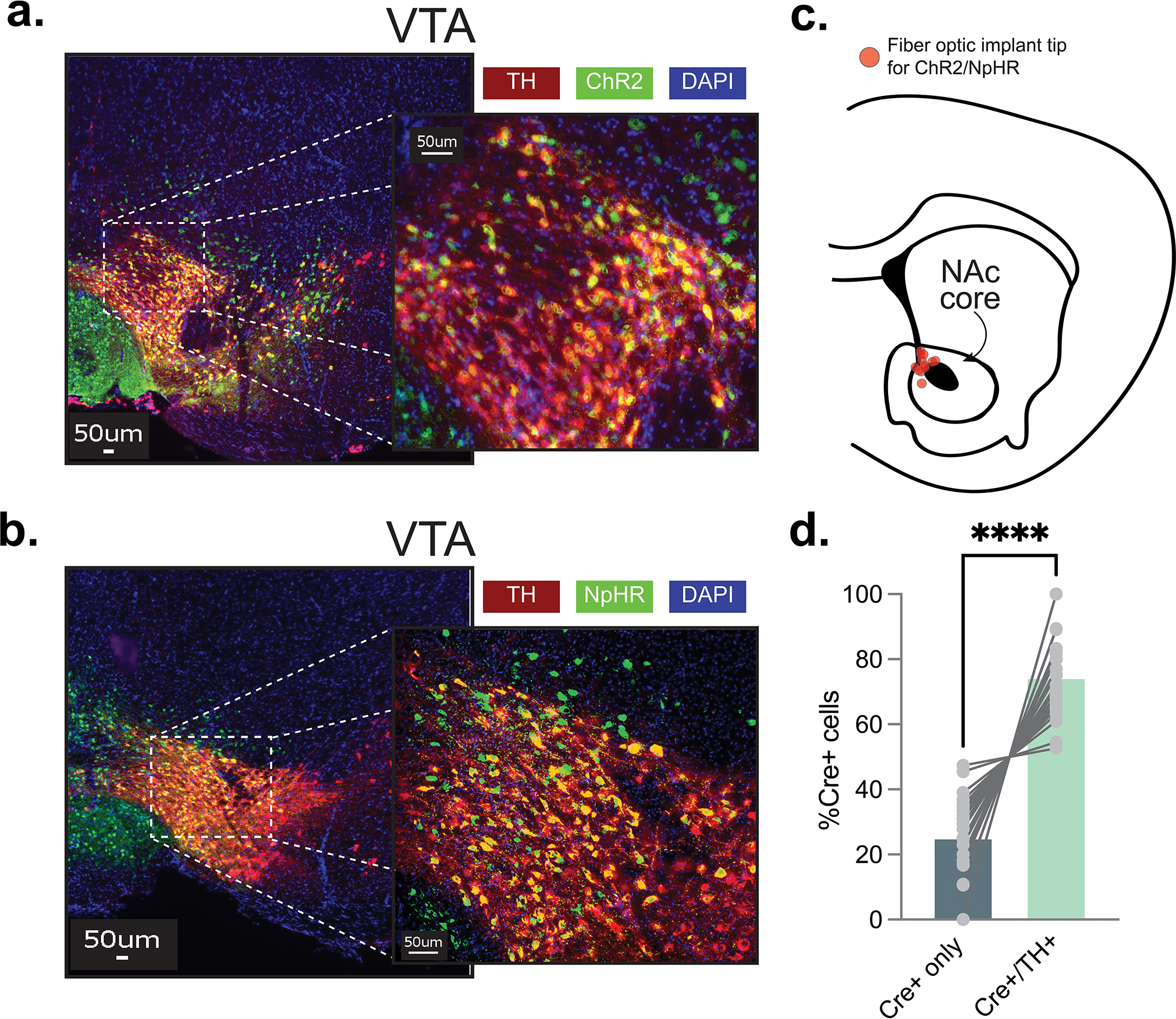
Extended Data Fig. 7. Validation of TH+ cell-specific opsin expression.

Optogenetics studies were designed to test whether the latent inhibition effect is controlled by the NAc core dopamine response to the pre-exposed fear cue. (a) Representative images showing the expression of ChR2 and TH in the VTA dopamine cell bodies. AAV9.rTH.PI.Cre.SV40 and AAV5.Ef1a.DIO.hchR2.eYFP or AAV5.Ef1a.DIO.eYFP was injected into the VTA to achieve specific expression of Chr2 in dopamine neurons. Specifically, AAV9.rTH.PI.Cre.SV40 injections resulted in Cre expression in all Tyrosine Hydroxylase (TH) positive cells within the VTA. By placing a fiberoptic above the NAc core, we were able to stimulate dopamine release from VTA projecting dopamine terminals in the NAc core. (b) Representative images showing the expression of NpHR and TH in the VTA dopamine cell bodies using the same approach as described. AAV9.rTH.PI.Cre.SV40 and AAV5.hSyn.eNpHR.3.0.eYFP or AAV5.Ef1a.DIO.eYFP were injected into the VTA and a fiberoptic was placed in the NAc core. (c) Schematic showing histologically verified fiber optic placements for all mice (n= 21 mice, 9 males, 12 females). (d) Cell counts were completed within the VTA from the experiments using the TH-specific excitatory/inhibitory opsin strategy. About 75% of the Cre+ cells in the VTA were also TH+ suggesting a significant portion of the ChR2 and NpHR cells were dopaminergic (two-sided paired t-test t22= 8.96, p=0.00000001). Data represented as mean ± S.E.M., **** p < 0.0001.
