
FIG 6.
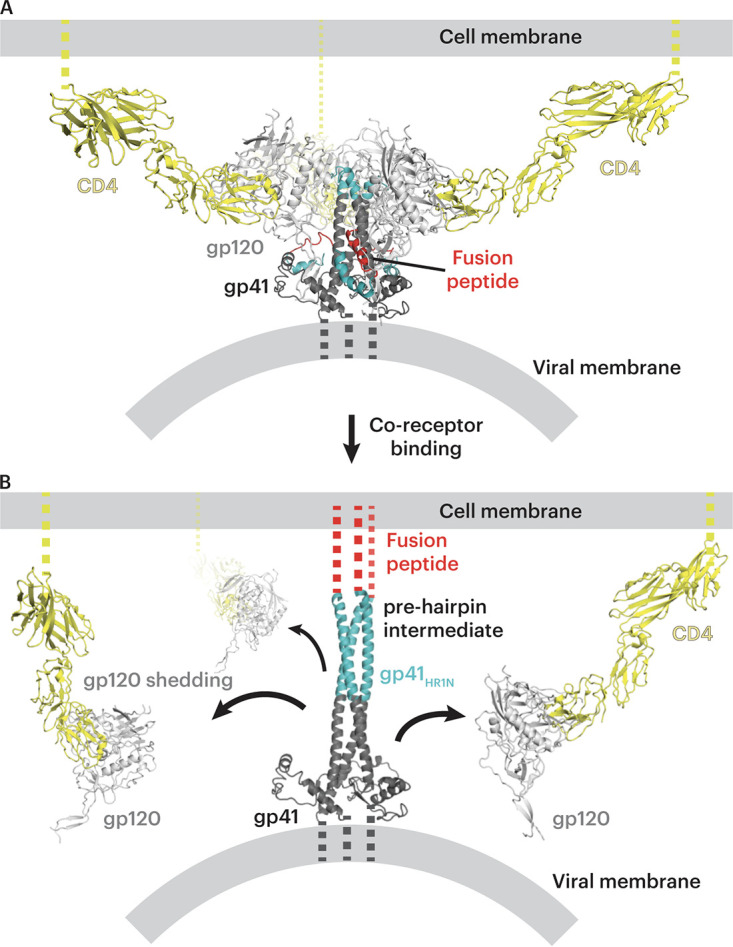
Model of postinfection formation of CD4-gp120 complexes on the host cell. (A) Open-conformation Env trimer (light and dark gray with fusion peptide in red and the N terminus of gp41 in cyan) on a viral membrane after binding to the host cell CD4 (yellow) on the host membrane. (B) Model of the prehairpin intermediate structure linking the viral and host cell membranes that is formed after host cell coreceptor binding (68). As potential targets to elicit V2i and CD4i antibodies during HIV-1 infection, cell surface CD4-gp120 complexes formed after gp120 shedding (black arrows) are shown on the host cell membrane.