. 2022 Nov 30;14(1):29–53. doi: 10.1039/d2sc04962g

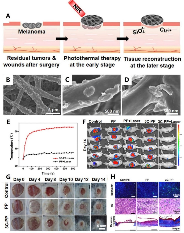
Fig. 2. (A) Schematic illustration of the CaCuSi4O10 nano-sized particle (NP)-coated fibrous scaffolds for photothermal therapy and tissue regeneration. (B–D) SEM images of the CaCuSi4O10 NP-coated fibrous scaffolds. (E) Temperature changes of the tumors treated with laser-irradiated PP and 3C-PP scaffolds (0.45 W cm−2, 15 min). (F) The whole-body fluorescence imaging of tumors on day 14 (n = 5). (G) Wound photographs at chronic wounds of different groups after fibrous scaffold implantation. (H) Immunofluorescence (green: CD31 and blue: nuclei), H&E staining and Masson's Trichrome staining images of skin wounds on day 14. Adapted with permission from ref. 96. Copyright 2018, Elsevier Ltd.

Fig. 2