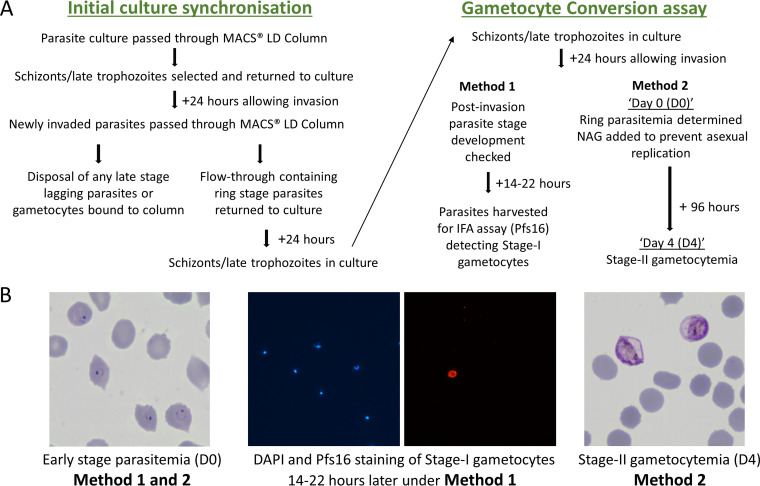
FIG 1.
Comparison of two different methods to assay P. falciparum gametocyte conversion rates in culture (details in Materials and Methods). (A) Scheme outlining the timing of processes undertaken for the different methods, Assay Method 1 involving staining of Pfs16 as a marker of early gametocytes, and Assay Method 2 involving the counting of stage-II gametocytes after 4 days of development in cultures treated with N-acetyl-d-glucosamine (NAG) to prevent asexual parasite replication. (B) Representative microscopy images of parasites on Giemsa-stained slides used for counting ring stage parasitemia early in the cycle (day 0); counting of proportions of stage-I gametocytes 16 to 22 h later by staining of the early gametocyte differentiation marker Pfs16 (DAPI staining of parasite DNA in blue, and anti-Pfs16 monoclonal antibody staining in red) for Assay Method 1; and counting stage-II gametocytemia in the NAG-treated culture after 96 h (day 4) for Assay Method 2.

