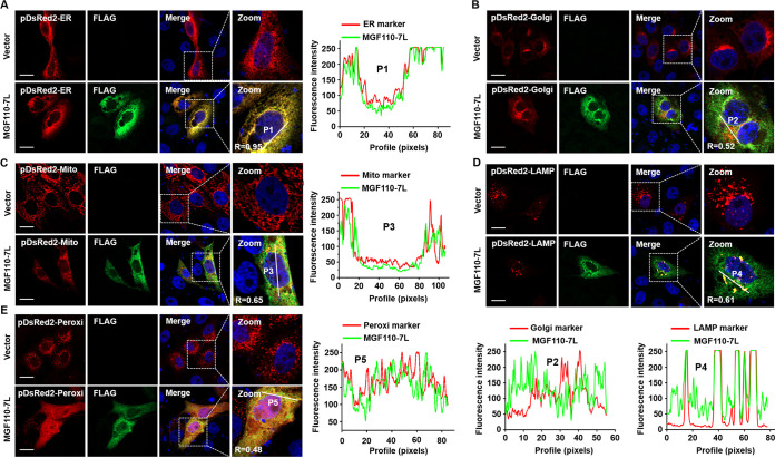
FIG 6.
Confocal immunofluorescence analysis of the intracellular distribution of ASFV MGF110-7L protein. PK-15 cells were cotransfected with an empty Flag vector or a MGF110-7L-Flag-expressing vector (0.5 μg) and one of the following organelle markers: pDsRed2-ER (A), pDsRed2-Golgi (B), pDsRed2-Mito (C), pDsRed2-LAMP1 (D), or pDsRed2-Peroxi (E) (0.2 μg). At 24 h posttransfection, the cells were fixed, permeabilized, and incubated first with anti-FLAG antibody and then with secondary antibody conjugated with AF488 (green). Nuclei were counterstained with DAPI (blue). The organelle marker was directly visualized (red), and localization was determined using confocal microscopy. The overlapping coefficient (R) was shown in enlarged images, and the intensity profile of the linear region of interest (ROI) across the PK-15 cell contained with MGF110-7L and the indicated organelle markers. Scale bar, 20 μm.

