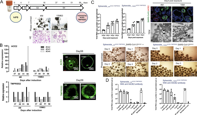
FIG 1.
Induction of Spheroids_4MACE-TMPRSS2 and Spheroids_15M63ACE-TMPRSS2 from human induced pluripotent stem cells. To obtain large amounts of three-dimensional (3-D) spheroids for anti-SARS-CoV-2 assays, two hiPSC lines, TkDN4-M and 15M63, were differentiated in suspension culture using bioreactors. These hiPS cell lines were established from different donors. After confirming the expression levels of ACE2 and TMPRSS2, key factors for SARS-CoV-2 infection, the induced SpheroidsACE-TMPRSS2 (Spheroids_4MACE-TMPRSS2 and Spheroids_15M63ACE-TMPRSS2) were examined on day 58 postinduction for susceptibility to SARS-CoV-2 infection and the effect of anti-human ACE2 monoclonal antibody on their susceptibility to SARS-CoV-2. (A) Overview of the 4-stage differentiation protocol of hiPSC lines in suspension culture, and representative phase-contrast and hematoxylin-and-eosin (HE)-staining images of hiPSC-derived spheroids on day 58. S, stage. (B) Left, the mRNA expression levels of ACE2 and TMPRSS2 relative to the levels in the control (human lung tissue) were examined using RT-qPCR; right, immunocytostaining (in green) was performed for detecting ACE2 and TMPRSS2 protein expression levels. #342, #343, and #344 indicate experiments 1, 2, and 3, respectively. N.D., not determined. (C) Left, time courses of SARS-CoV-2 infection and replication in SpheroidsACE-TMPRSS2S were examined with RT-qPCR targeting the SARS-CoV-2 nucleocapsid. Bars and error bars show geometric mean values ± standard deviations (SD); points represent data from individual microplate wells (n = 6). Right and bottom, representative morphological images from six independently conducted experiments are shown. Right, immunocytostaining to detect SARS-CoV-2 protein with a convalescent IgG fraction that was isolated from serum of a convalescent COVID-19 individual (top) (SARS-CoV-2 antigens, ACE2, and nuclei are indicated in green, red, and blue, respectively) and TEM observation (bottom) (arrows indicate virus particles) were conducted. Bottom, no cytopathic effect on either infected spheroid line was observed with phase-contrast microscopy. (D) Pretreatment and coincubation with anti-human ACE2 MAb with SpheroidsACE-TMPRSS2S blocked SARS-CoV-2 infection. Bars and error bars show geometric mean values ± SD; points represent data from individual microplate wells (n = 3). Details of the methods are described in the supplemental material.

