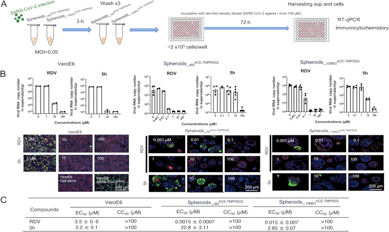
FIG 2.
Evaluation of anti-SARS-CoV-2 agents using SpheroidsACE-TMPRSS2. The two induced spheroid lines, Spheroids_4MACE-TMPRSS2 and Spheroids_15M63ACE-TMPRSS, were exposed to SARS-CoV-2WK521 at a multiplicity of infection (MOI) of 0.05. Vero E6 cells were also exposed to SARS-CoV-2WK-521 (MOI of 0.05) as a control experiment. Three hours postexposure, the virus was washed out three times with culture medium and the cells seeded in 96-well microtiter culture plates at a density of 2 × 10⁶ cells/well and incubated with or without test compounds for 72 h. On day 3 postexposure, culture supernatants were collected for quantitative SARS-CoV-2 RNA analysis using RT-qPCR, while cells were fixed and subjected to quantitative analysis using immunocytostaining. EC50 values were calculated with the obtained data using RT-qPCR. (A) Scheme of the experimental setup. sup, supernatant. (B) Top, inhibition of SARS-CoV-2 infection in SpheroidsACE-TMPRSS2S and Vero E6 cells was examined with RT-qPCR. Bars and error bars show geometric mean values ± SD; points represent data from individual microplate wells (n = 4). Bottom, representative immunocytostaining images from four independently conducted experiments are shown. SARS-CoV-2 antigens, F-actin, and nuclei are indicated in green, red, and blue, respectively. (C) EC50 values and CC50 values of anti-SARS-CoV-2 agents in SpheroidsACE-TMPRSS2S were determined using RNA-qPCR and the water-soluble tetrazolium salt (WST-8) based cell viability assay, respectively. Data are mean values ± SD (n = 4). Details of the methods are described in the supplemental material.

