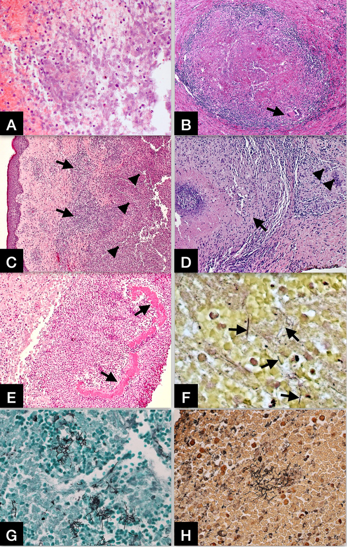
FIG 1.
(A) Nocardial brain abscess. Routine hematoxylin & eosin (H&E) stain shows necrosis, karyorrhectic debris, and neutrophilic infiltrate in the abscess. Original magnifications: X200. (B) Nocardial lung infection with granulomatous inflammation. H&E stain shows a granuloma with central necrosis, lymphohistiocytic infiltrate at the periphery, and rare multinucleated cells (arrow). Original magnifications: X100. (C) Nocardial skin infection. H&E stain shows a zone of mixed inflammatory infiltrate (arrows) and large area of abscess formation (arrowheads) in the dermis. Original magnifications: X50. (D) Nocardial skin infection with granulomatous inflammation. H&E stain shows a granuloma (arrow) with central necrosis and lymphohistiocytic infiltrate at the periphery, as well as scattered multinucleated cells in adjacent area (arrowheads). Original magnifications: X100. (E) Nocardial skin infection. H&E stain shows dense aggregated granules (grains) with a peripheral radial deposition of intensely eosinophilic material (arrows) – a Splendore-Hoeppli phenomenon. Original magnifications: X100. (F) Gram stain showing scattered Gram-positive and gram-variable nocardial organisms in the abscess. Original magnifications: X400. (G) Grocott methenamine silver (GMS) stain showing clusters of filamentous nocardial organisms in the abscess. Original magnifications: X400. (H) Steiner silver stain showing clusters of filamentous nocardial organisms in the abscess. Original magnifications: X400.

