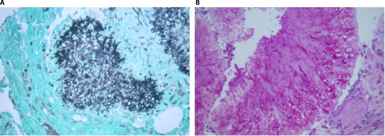
FIG 4.
Hyalohyphomycosis-related osteomyelitis. Shown is the histopathology of the left lateral malleolus depicted in Fig. 6 in a patient with osteomyelitis caused by Scedosporium spp. (A) Gomori methenamine-stained fungal balls and fungal hyphae branched at 45° invading bone tissue (magnification, ×400). (B) Periodic acid-Schiff-stained fungal balls with peripheral zonation and septate hyphae (magnification, ×400).

