FIG 8.

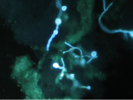
Phaeohyphomycotic osteomyelitis. Shown are hyphae with intercalary and terminal chlamydoconidium swellings as seen by the direct preparation of infected tissue using Blankophor P fluorescent stain (magnification, ×400).

Phaeohyphomycotic osteomyelitis. Shown are hyphae with intercalary and terminal chlamydoconidium swellings as seen by the direct preparation of infected tissue using Blankophor P fluorescent stain (magnification, ×400).