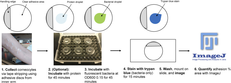
FIG 1.
Schematic of the corneocyte adhesion assay. (Step 1) Corneocytes are collected with tape stripping discs from the upper or lower arm near the elbow of a volunteer with healthy skin. (Step 2) Corneocytes can be preincubated with protein for 45 min or (step 3) incubated with fluorescent bacteria at an OD600 of 0.15 for 45 min. (Step 4) Corneocytes incubated with bacteria only are stained with trypan blue for 15 min. (Step 5) After incubation and/or staining, corneocytes are washed, mounted on a slide, and imaged with a fluorescence microscope. (Step 6) Bacterial images are quantified for percent area of adhesion with Fiji ImageJ.

