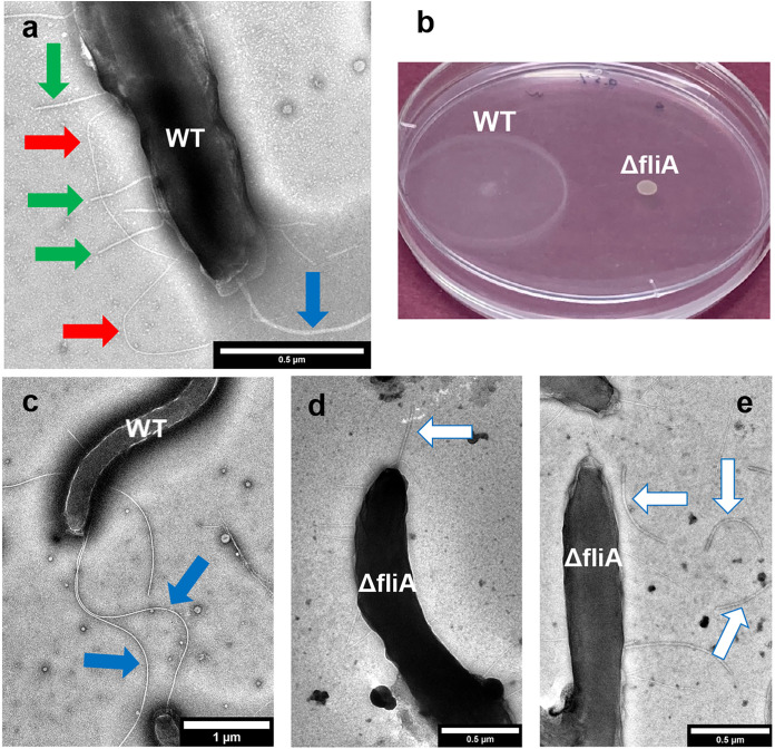
FIG 2.
D. ferrophilus filament expression and motility. (a and c) Transmission electron micrographs of a typical wild-type (WT) cell. Colored arrows highlight features as follows: blue arrow, putative flagella; red arrow, long flexible putative pili; green arrow, short, straight putative pili. (b) Motility assay of wild-type (WT) and ΔfliA strains. One of duplicate plates (0.5% agar) is shown. Similar results were observed with an 0.7% agar concentration (data not shown). (d and e) Transmission electron micrographs of ΔfliA strain cells. White arrows highlight truncated flagella, some of which are disassociated from the cells.

