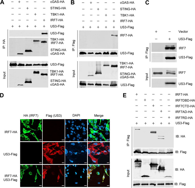
FIG 5.
DEV US3 interacts with the activation domain of IRF7. (A and B) DEFs were transfected with the indicated plasmids for 36 h before coimmunoprecipitation and immunoblot analyses with the indicated antibodies. (C) DEFs were transfected with a US3-Flag expression plasmid or an empty vector; 36 h posttransfection, a coimmunoprecipitation assay was performed with anti-Flag antibody. (D) DEF cells were transfected with US3-Flag and/or IRF7-HA expression plasmids for 24 h and then fixed and processed for dual labeling. Cell nuclei were counterstained with DAPI (blue). US3 (red) and IRF7 (green) proteins were visualized by immunostaining with anti-Flag and anti-HA antibodies, respectively. Bars, 10 μm. (E) A US3-Flag expression plasmid was cotransfected with IRF7 (aa 1 to 512) or a series of truncation mutants of IRF7, including IRF7DBD (aa 1 to 141), IRF7CTD (aa 142 to 512), IRF7AD (aa 142 to 320), and IRF7IRD (aa 321 to 512), into HEK293T cells; 36 h posttransfection, the cell lysates were immunoprecipitated with anti-Flag antibodies and analyzed by Western blotting.

