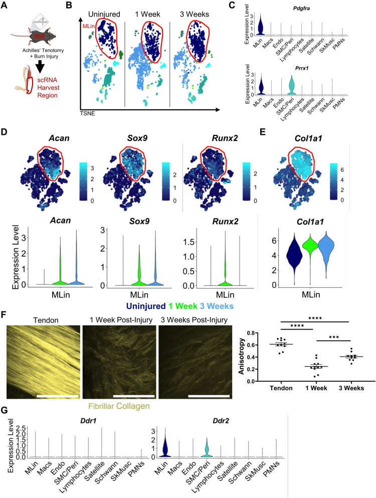
Fig. 1. MLin cells express fibrillar collagen and collagen binding protein DDR2 after injury.
(A) Schematic of BT injury and single-cell harvest region. (B) TSNE plot of scRNA sequencing of cells harvested from uninjured and 1 and 3 weeks following injury highlighting MLin population in red. (C) Violin plot of relative expression of genes encoding MLin markers Pdgfra and Prrx1. (D) TSNE and violin plots of relative expression of chondrogenic genes (Acan and Sox9), osteogenic gene Runx2, and (E) Col1a1. (F) Second harmonic generation (SHG) imaging of fibrillar collagen in uninjured tendon and at the injury site 1 and 3 weeks following injury. Graph showing anisotropy quantification of collagen ECM (n = 3 mice per group, one to two images per mouse, bars are means ± SEM). Scale bars, 100 μm. (G) Violin plots of relative expression of DDR1 and DDR2 genes Ddr1 and Ddr2. Plots include all time points.

