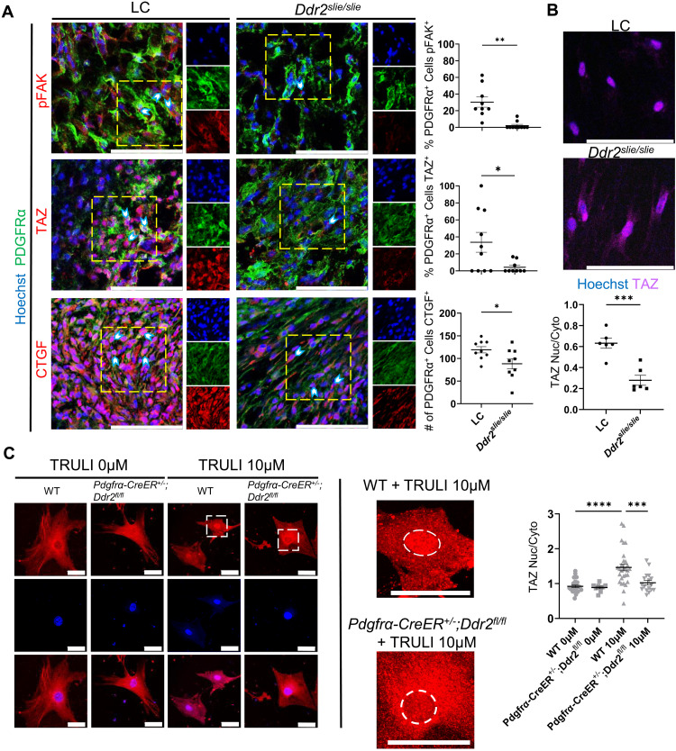
Fig. 6. DDR2 deletion down-regulates the FAK/YAP/TAZ axis following injury.
(A) Representative confocal microscope images (63×) of injured hindlimb of LC and Ddr2slie/slie mice 1 week after BT, stained with Hoechst, anti-PDGFRα, and anti-phosphorylated FAK (pFAK), anti-TAZ, or anti-CTGF with quantification of the percentage or number of PDGFRα-expressing cells with positive staining of each antigen of interest (n = 3 mice per group, n = 2 to 4 images per mouse). Scale bars, 100 μm. (B) Two-dimensional aligned collagen plates with MLin cells from Ddr2slie/slie uninjured hindlimb and LC uninjured hindlimb and stained with Hoechst and TAZ. Graph shows ratios of positive TAZ staining localized to the nucleus compared to TAZ staining in the cytoplasm of cells from Ddr2slie/slie uninjured hindlimb and LC uninjured hindlimb (n = 2 mice per group, n = 3 images per mouse, bars are means ± SEM). Scale bars, 50 μm. (C) Representative confocal images (40×) of MLin cells isolated from Pdgfrα-CreER+/−;Ddr2fl/fl and wild-type mice 1 week after BT plated on three-dimensional collagen plates treated with 0 or 10 μM TRULI. Cells were stained with Hoechst and anti-TAZ (n = 19 to 29 cells per sample). Scale bars, 100 μm. The region within white dashed lines of high-magnification images is the nucleus. All graphs show means ± SEM.

