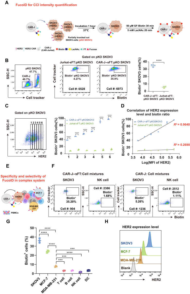
Fig. 4. Characterization of the selectivity and specificity of CAR-J–sFT probe in CAR-J–SKOV3 interaction system.
(A) Workflow of analyzing CCI intensity in the model of CAR-antigen–mediated interactions. (B) Representative flow cytometric plots and statistics showing biotinylation of pKO SKOV3 cells mediated by CAR-J–sFT and Jurkat-sFT. n = 3. The background is defined as the signal produced on pKO SKOV3 incubated with Jurkat without membrane-anchored sFT. (C) Statistical analysis of labeling ratio among six groups of pKO SKOV3 cells with different HER2 expressions. (D) Correlation analysis of biotin ratio and HER2 expression level in the six groups divided in (C). (E) Schematic illustration of CAR-J–sFT as a probe for detecting the specificity and selectivity of FucoID in a complex system. (F and G) Flow cytometry–based quantification of selective and specific labeling of SKOV3, MCF-7, and MDA-MB-231 in cell mixtures by CAR-J–sFT (doped with SKOV3, MCF-7, and MDA-MB-231 in PBMCs at a ratio of 1:1:1:10). n = 3. NK, natural killer. (H) Flow cytometric analysis of expression level of HER2 on SKOV3, MCF-7, and MDA-MB-231. ***P < 0.001; ****P < 0.0001.

