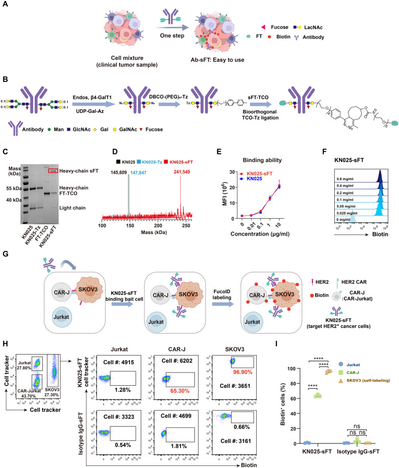
Fig. 6. Development of Ab-sFT for detecting CCIs.
(A) Schematic illustration of the application of Ab-sFT probes to study clinical samples. (B) Characterization of synthesis process of Ab-sFT. (C and D) Characterization of KN025, KN025-Tz, and KN025-sFT by (C) SDS-PAGE and (D) mass spectrometry. (E) Titration of the HER2 binding ability of KN025-sFT. (F) Characterization of FT activity of KN025-sFT by labeling LacNAc on NK92 cells with GF-Biotin. (G) Schematic overview of probing the interactions between CAR-J/Jurkat-SKOV3 by KN025-sFT. CAR-J, Jurkat, and SKOV3 were mixed as a model of CCI. KN025-sFT was added to the system to label HER2+ cells and their interacting cells. (H) Flow cytometric analysis and (I) summary statistics of the interaction-dependent fucosyl-biotinylation of Jurkat, CAR-J, and SKOV3 in the model system. n = 3. Interaction time, 1 hour; labeling time, 30 min. The background is defined as the signal produced on cells treated with anti-HER2 antibody. ns, P > 0.05; ****P < 0.0001.

