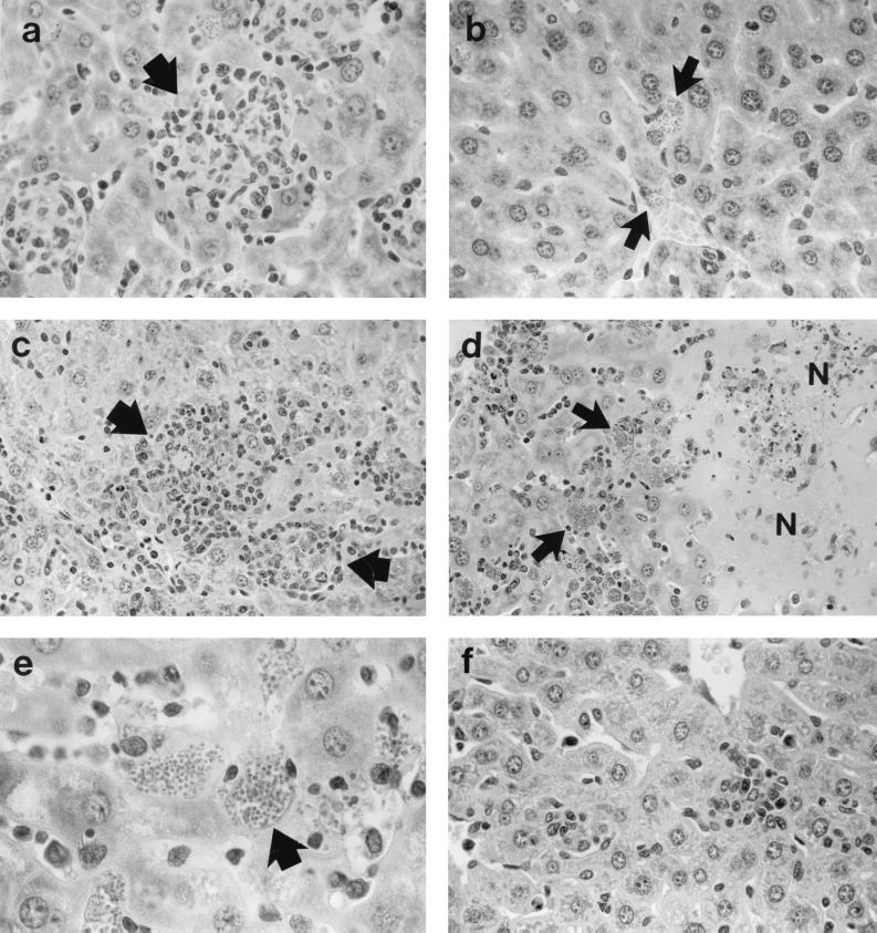
FIG. 2.
Liver histologic response to L. donovani 2 to 4 weeks after infection in WT controls (a) and TNF KO mice (b to f). (a and b) Week 2. WT mice (a) show normal developing granulomas (arrow) at sites of parasitized Kupffer cells; in contrast, in KO mice (b), there is no cellular response at parasitized foci (arrows). (c) Week 3. Rapid granuloma development in KO mice. (d and e) Week 4. KO mice show destructive inflammation with few recognizable granulomas, widespread areas of necrosis (N), and abundant cellular debris (d). Necrotic areas in panel d are ringed by partially preserved tissue containing large masses of intracellular amastigotes (arrows), also shown in panel e. (f) Week 3. Near absence of inflammatory changes in KO mice treated with AmB during the previous week. Magnifications, ×315 (a, b, and f), ×200 (c and d), and ×500 (e).

