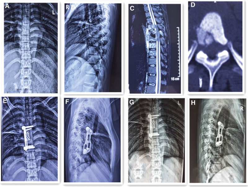
Figure 2.
Fifty-six-year-old man with short segment thoracic vertebra tuberculosis (T6-T7 level). (A, B) Preoperative frontal and lateral x-rays showed bone destruction in the 6/7 vertebral body and narrowing of the vertebral space, with a kyphosis angle of 15°. (C) Preoperative magnetic resonance imaging scan shows vertebral body destruction and abscess. (D) Preoperative computed tomography scans demonstrate vertebral body destruction. (E, F) Radiographs postoperatively showing well-positioned internal fixation. (G, H) Radiographs showing satisfactory focal clearance and strut graft stability at the final follow-up.

