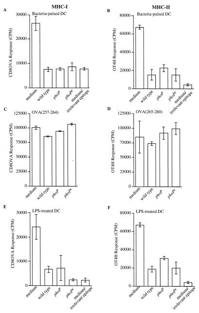
FIG. 1.
Reduced presentation of bacterial antigens on MHC-I and MHC-II molecules by DC exposed to serovar Typhimurium or LPS with modified lipid A. DC were pulsed with bacteria for 2 h and after washing and gentamicin treatment were incubated for another 22 h (A to D), or DC were treated with purified LPS for 24 h (E and F). The DC were then pulsed with E. coli expressing Crl-OVA or E. coli expressing Crl-HEL (containing an epitope irrelevant for the T-cell hybridoma [medium/irrelevant epitope]) (A, B, E, and F), OVA(257-264) peptide (C), or OVA(265-280) peptide (D) for 2 h. Following paraformaldehyde fixation, OVA(257-264)/Kb (A, C, and E) or OVA(265-277)/I-Ab presentation (B, D, and F) was quantitated by adding CD8OVA or OT4H T-hybridoma cells, respectively. (A to D) The x axis indicates that the DC were incubated in medium alone during the initial 2 h (medium or medium/irrelevant epitope) or were pulsed for 2 h with bacterial strains as follows: wild type, serovar Typhimurium ATCC 14028; phoP, serovar Typhimurium CS015; phoPc, serovar Typhimurium CS022. (E and F) The x axis indicates that the DC were incubated in medium alone for 24 h (medium or medium/irrelevant epitope) or indicates the type of purified LPS (1 μg/ml) present during the initial 24-h incubation: wild type, LPS purified from serovar Typhimurium ATCC 14028; phoP, LPS purified from serovar Typhimurium CS015; phoPc, LPS purified from serovar Typhimurium CS022. Data are presented as the means of triplicate samples ± 1 standard deviation. Maximum CTLL proliferation after exposure to recombinant IL-2 was 160,000 cpm. Similar results were obtained in at least three independent experiments.

