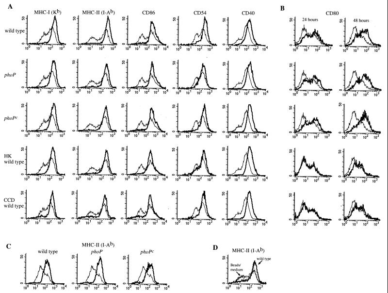
FIG. 2.
DC coincubation with serovar Typhimurium alters expression of surface molecules important in signaling the immune system. (A and B) DC were coincubated with either viable wild-type, phoP, or phoPc serovar Typhimurium; viable wild-type serovar Typhimurium in the presence of CCD; or heat-killed (HK) wild-type serovar Typhimurium, as indicated to the left of each row of histograms. After an initial 2-h pulse with bacteria, the cells were washed, treated with gentamicin, and incubated an additional 22 h (A and B) or 46 h (B) before flow cytometry was performed. (C) DC were coincubated with LPS purified from either wild-type, phoP, or phoPc serovar Typhimurium as indicated. The surface expression of MHC-II molecules on DC after 24 h of stimulation with LPS (thick line) compared to that on DC incubated in medium only (thin line) is shown. (D) The surface expression of MHC-II molecules on DC 48 h after the addition of latex beads (dotted line) compared to that on DC incubated in medium only (thin line) or DC incubated with wild-type serovar Typhimurium (thick line) is shown. The upregulation of the different surface markers was not due to unspecific binding of Ig used for the fluorescence-activated cell sorter analysis, as appropriate Ig isotype subclass controls showed no difference in expression levels for infected and uninfected cells (not shown). Similar results were obtained in at least four independent experiments.

