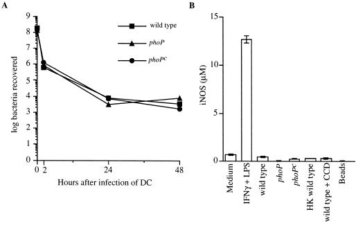
FIG. 6.
DC control intracellular replication of serovar Typhimurium within a 48-h period. (A) Following a 2-h pulse with either wild-type, phoP, or phoPc serovar Typhimurium, DC were washed and were either lysed to determine the number of bacteria recovered at the 2-h time point or treated with gentamicin before continuing the incubation for a total of 24 or 48 h. At these time points, DC were lysed and the number of viable bacteria remaining was determined by plating on LB agar plates. The actual initial bacterium-to-DC infection ratios were 15:1, 13:1, and 17:1 for wild-type, phoP, and phoPc bacteria, respectively, as determined by viable counts. (B) NO2− was quantitated in the supernatants of DC that were incubated in medium alone or in medium containing IFN-γ (300 U/ml) and LPS (10 μg/ml); or infected with wild-type, phoP, or phoPc serovar Typhimurium or with wild-type serovar Typhimurium in the presence of CCD or with heat-killed (HK) wild-type serovar Typhimurium; or incubated with 1-μm polystyrene beads, as indicated. DC were pulsed with bacteria for 2 h. The cells were washed and treated with gentamicin, and incubation was continued for an additional 46 h. At this time point the level of NO2− in the culture supernatant was quantitated by using the method of Greiss with NaNO2 as the standard. Data are presented as the means of triplicate samples ± 1 standard deviation. Similar results were obtained in at least three independent experiments.

