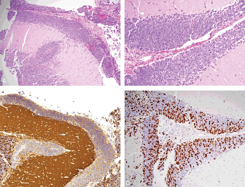
Fig. 4.

Histological and immunohistochemical specimens. The hematoxylin and eosin-stained section ( A ) showed the neoplastic infiltrate within the subarachnoid space (original magnification 100 × ). A higher magnification ( B ) revealed the neoplastic cells with large, round-oval nuclei, and scant cytoplasm (original magnification 400 × ). The immunohistochemical staining documented the positivity for synaptophysin ( C ) and a high proliferation index ( D ).
