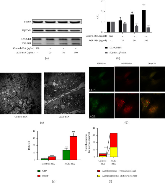
Figure 3.

AGE-BSA induced retinal pericytes autophagy. (a, b) The expression of LC3A/B and SQSTM1 in retinal pericytes were measured by the western blot. (c) Transmission electron microscopy images of retinal pericytes, the arrows indicated the autophagosomes. Scale bar = 2 μm. (d) The immunofluorescence assays were performed in retinal pericytes, which were transfected with GFP-mRFP-LC3 adenovirus. Scale bar = 50 μm. (e) The mean number of GFP and mRFP dots/cell. (f) The yellow dots represent the average number of autophagosomes in merged images per cell and the red dots represent the average number of autolysosomes in merged images per cell. ∗∗∗p < 0.001 vs control group, ∗∗p < 0.01 vs control group, and ∗p < 0.05 vs control group; p values were calculated using Turkey's t-test; A. U., arbitrary units.
