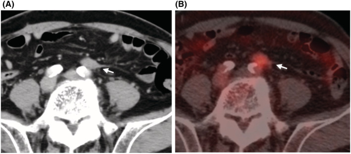
FIGURE 1.

CECT scan and 18F‐FDG‐PET/CT findings in Case1. CECT scan indicates a tumor formation (21 × 12 mm) on the caudal ventral side of the left common iliac artery bifurcation (white arrow, A), corresponding to the SUVmax 5.2 tumor in the 18F‐FDG‐PET/CT scan (white arrow, B).
