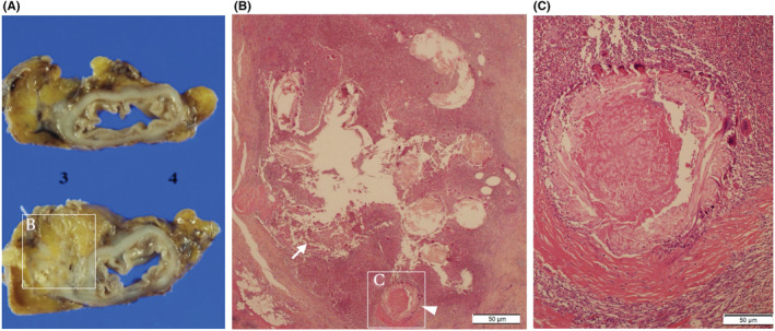
FIGURE 4.

Macroscopic and microscopic findings in Case2. Resected specimen revealed that the tumor was an ill‐defined hard elastic mass, and its size was 50 × 70 × 25 mm (A). Pathological examination reveals an abscess formation (white arrow) was observed (B) around the surgical threads (arrowhead) in the submucosa (C). No neoplastic lesions were identified.
