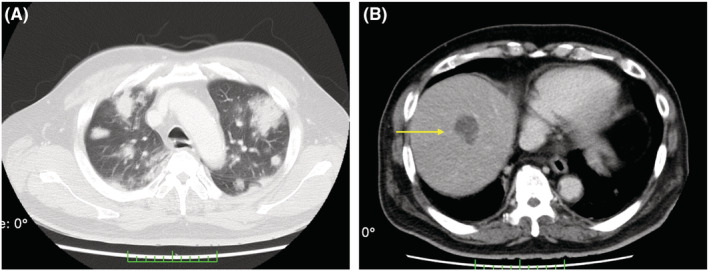
FIGURE 2.

CT scan without contrast‐enhancement on the 3rd hospital day showing cotton‐like shadow and rounded infiltrate with air bronchogram; the margin of the infiltrate is fluffy, and the inside is granular in the lung (A); the liver abscess is enlarged to 33 mm on CT scan with contrast‐enhancement (B).
