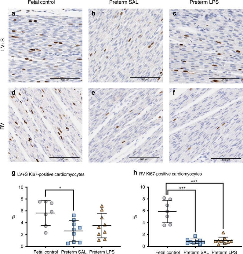
Fig. 3. Ki67 immunostaining of proliferating cardiomyocytes.
Representative images of Ki67 immunohistochemical staining in the left ventricle + septum (LV + S; a–c) and right ventricle (RV; d–f) from aged-matched fetal lambs (fetal control, n = 7) and preterm lambs exposed antenatally to saline (preterm SAL, n = 9) or LPS (preterm LPS, n = 9). Scale bar = 100 μm. Graphs show the proportion of Ki67 positively-labeled cardiomyocytes in the LV + S (g) and RV (h) of each experimental group (gray circles = fetal control; blue squares = preterm SAL; orange triangles = preterm LPS). Statistical analyses were performed using a one-way ANOVA followed by a Bonferroni post hoc test. Data are presented as mean ± SD, * denotes p < 0.05, *** denotes p < 0.001.

