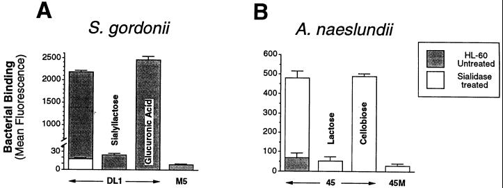
FIG. 4.
Flow cytometric analysis of the specificity of bacterial binding to HL-60 cells incubated with DMSO for 5 days. (A) Binding of fluoresceinated S. gordonii DL1 and M5. (B) Binding of fluoresceinated A. naeslundii WVU45 and WVU45M. HL-60 cells were untreated or treated with sialidase. Where indicated, binding was performed in the presence of 10 mM sialyllactose, 10 mM glucuronic acid, 50 mM lactose, or 50 mM cellobiose.

