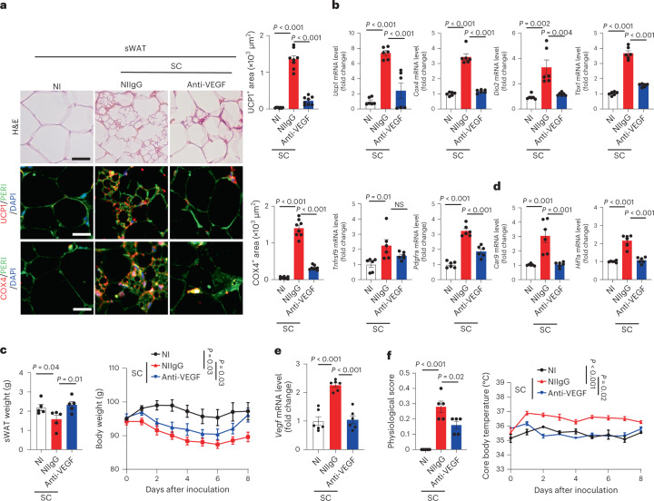
Fig. 3. SARS-CoV-2 induces VEGF-dependent adipose browning in hamsters.
a, Histological and immunohistochemical analyses of sWAT of NI hamsters and NIIgG- or anti-VEGF-treated day-8 post-SC-infected hamsters by staining with H&E, PERI, UCP1 and COX4. Tissue sections were counterstained with DAPI (blue). Quantification of UCP1- and COX4-positive signals in sWAT (n = 8 random fields per group). b, mRNA levels of browning markers of Ucp1, Cox4, Dio2, Tbx1, Tnfrsf9 and Pdgfra in sWAT of NI hamsters and NIIgG- or anti-VEGF-treated day-8 post-SC-infected hamsters were quantified by qPCR (n = 6 samples per group). c, sWAT weight and body weight of NI hamsters and NIIgG- or anti-VEGF-treated day-8 post-SC-infected hamsters (n = 5 hamsters per group). Statistics are shown on day 8 of infection. d, qPCR quantification of Car9 and Hif1a mRNA levels in sWAT of NI hamsters and NIIgG- or anti-VEGF-treated day-8 post-SC-infected hamsters (n = 6 samples per group). e, qPCR quantification of Vegf mRNA levels in sWAT of NI hamsters and NIIgG- or anti-VEGF-treated day-8 post-SC-infected hamsters (n = 6 samples per group). f, Physiological scores and core body temperature of NI hamsters and NIIgG- or anti-VEGF-treated day-8 post-SC-infected hamsters (n = 5 hamsters per group). Statistics are shown on day 8 of infection. Data are presented as mean ± s.e.m. Statistical analysis was performed using two-sided unpaired Student’s t-tests. Scale bar, 50 μm.

