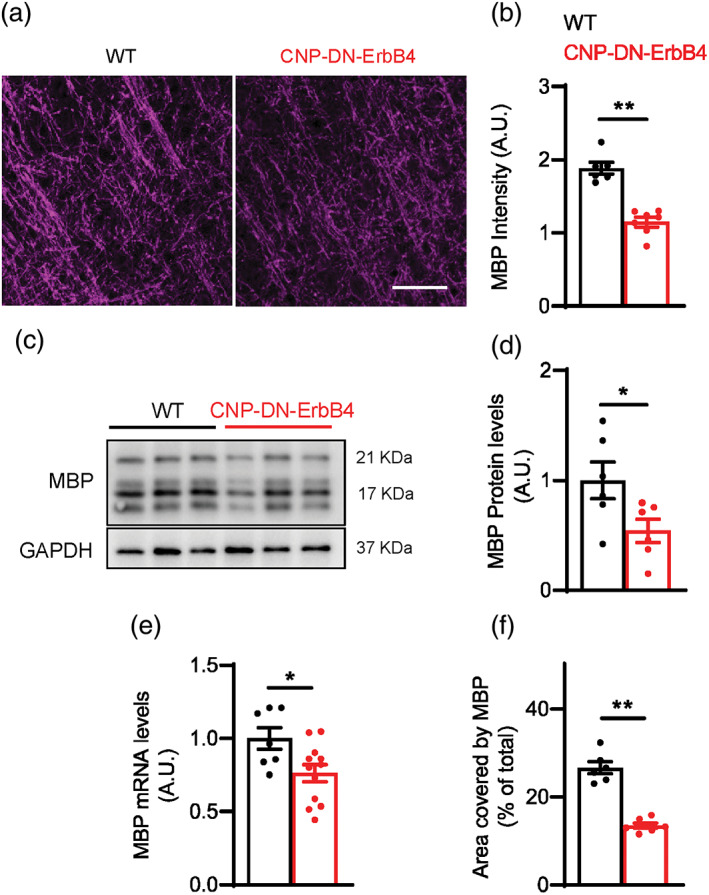
FIGURE 1.

Loss of oligodendrocyte ErbB receptor signaling leads to a reduction in MBP protein and mRNA expression levels in A1. (a) Representative photomicrographs of primary auditory cortex of WT and CNP‐DN‐ErbB4 mice showing the expression of myelin basic protein (MBP) (magenta). Scale bar: 20 μm. (b) Quantification of MBP staining intensity (WT: black; CNP‐DN‐ErbB4: red) (n = 6–7 mice per genotype; **p = .001). (c) Representative MBP and GAPDH Western blots of A1 samples from WT and CNP‐DN‐ErbB4 mice. (d) Quantification of MBP protein expression normalized by GAPDH in A1 of WT and CNP‐DN‐ErbB4 mice (n = 6; p = .0413). (e) MBP mRNA levels in A1 of WT and CNP‐DN‐ErbB4 mice (n = 7–12; p = .0226). (f) Quantification of the percent of area covered by MBP (WT: black; CNP‐DN‐ErbB4: red) (n = 6–7 mice per genotype; **p ˂ .001) (A.U.: arbitrary units). Unpaired two‐tailed Student's t test was performed. Data are expressed as mean ± SEM.
