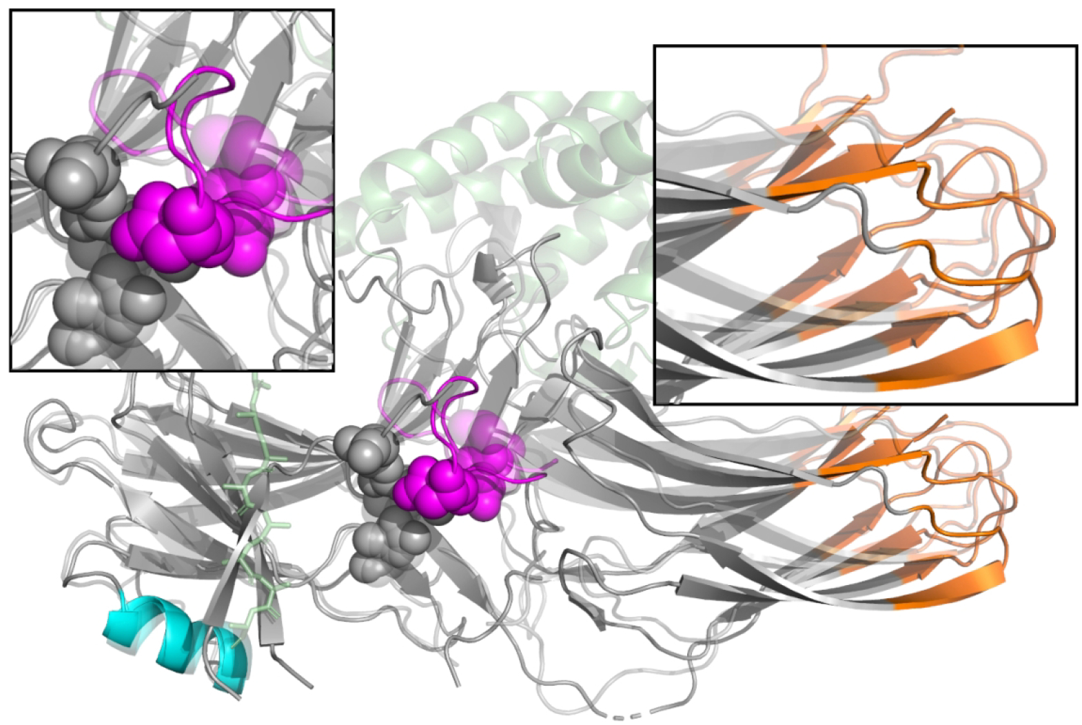
Figure 2:

Changes in the structure of arrestin upon activation. Inactive arrestin (solid) and active arrestin (translucent) are superimposed on touch of each other, with the N-terminal domain on the left as in Figure 1. The polar core (spheres), lariat loop (purple), GPCR (green), and GPCR C-terminal peptide (green sticks) are shown. Blue and purple regions represent sequence which is included in our ANM modelling. Arrestin activation is marked by the disruption of the polar core residues (top left panel), and a rotation of the C-domain with respect to the N-domain (top right panel).
