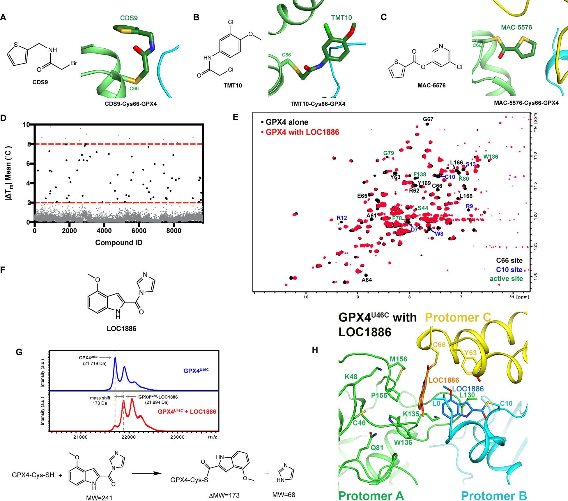
Figure 5. Screening of Lead-Optimized-Compound library identified lead compound binding to the Cys66 allosteric site.

A-C, Crystal structure of GPX4U46C with CDS9, TMT10, or MAC5576. D, Thermal shift assay was applied to screen 9,719 compounds in the Lead-Optimized-Compound library for in vitro binders of GPX4U46C. E, Overlap of 1H, 15N-HSQC-NMR spectrum of 50 μM 15N-GPX4U46C alone and its mixture with 800 μM LOC1886. F, Structure of LOC1886. G, Intact protein MALDI MS analysis of GPX4U46C preincubated with DMSO or LOC1886 and the proposed nucleophilic substitution reaction based on the observed mass shift. H, Co-crystal structure of GPX4U46C with LOC1886. The LOC1886 molecules bound to cysteine 66 (yellow) and cysteine 10 (cyan) of GPX4U46C are shown in orange and marine, respectively. The three protomers (A-C) are colored light-green, cyan, and yellow, and residues interacting with two LOC1886 molecules are shown as stick models and labeled.
See also Figure S5.
