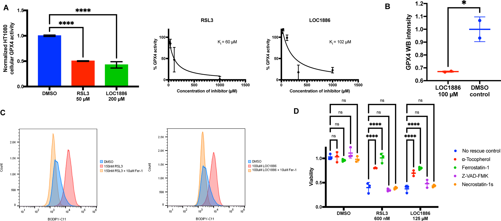
Figure 6. LOC1886 inhibits and degrades GPX4 to induce ferroptosis.

A, Left: Enzymatic activity of GPX4 in HT1080 cell lysates preincubated with DMSO, 50 μM RSL3, or 200 μM LOC1886 was acquired (n=10). One-way ANOVA followed by Dunnett’s multiple comparisons test was performed: p**** < 0.0001. Right: Measurement of inhibition constants (Ki) of RSL3 and LOC1886 on purified GPX4U46C protein (n=2). B, Native GPX4 in HT-1080 cells were tested for vulnerability to degradation induced by 100 μM LOC1886 (n=2). Unpaired t test was performed: p* < 0.05. C, Lipid peroxidation in HT-1080 cells treated with DMSO, 200 nM RSL3, or 100 μM LOC1886, with or without fer-1 rescue, was assayed by flow cytometry using C11-BODIPY. D, Viability of HT1080 cells treated with DMSO, 600 nM RSL3, or 125 μM LOC1886, with or without a-Tocopherol (100 μM), Ferrostatin-1 (100 μM), Z-VAD-FMK (20 μM), and Necrostatin-1s (20 μM) rescue (n=3). Two-way ANOVA followed by Dunnett’s multiple comparisons test was performed: pns>0.05 and p**** < 0.0001.
For A, B, and D, data are represented as mean ± s.d. See also Figure S5.
