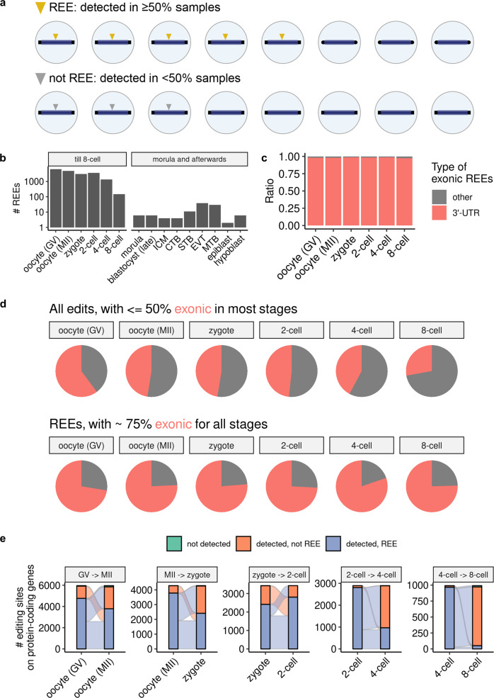
Fig. 2. Thousands of organized REEs were detected in early human embryos.
a Definition of REE. b Count of REEs from normal samples per stage. c Percentage of 3'-UTR REEs in early stages of embryogenesis. See also Supplementary Fig. 13 for percentages of general edits. d Percentage of exonic edits and REEs (shown in red) in the early stages of embryogenesis. e Sankey plot describing the numbers of REEs passed to subsequent early stages. For clarity, only REEs observed in at least one of the two stages appear in each subplot. For c–e, we considered only REEs on protein-coding genes. See also Supplementary Figs. 14 and 15 for the percentage of 3'-UTR REEs, and the percentage of exonic edits and REEs in the late stages of embryogenesis.

