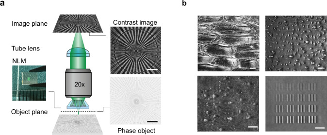
Fig. 1. A nonlocal metasurface (NLM) patterned on a microscope slide enables phase contrast imaging.
a A schematic showing how a classic bright field microscope can be transformed into a phase imaging setup simply by inserting an NLM on top of the phase object. The flat optical component, a commercial phase object and the corresponding phase contrast image are shown next to the microscope schematic. b NLM phase contrast images of onion cells (top left), human osteosarcoma cells (U-2 OS, top right), a dense array of exfoliated transparent hexagonal boron nitride (hBN) flakes (bottom left), and arrays of metasurface elements comprised of silicon nitride pillars with different diameters (bottom right). All scale bars in a and b correspond to 25 µm length.

