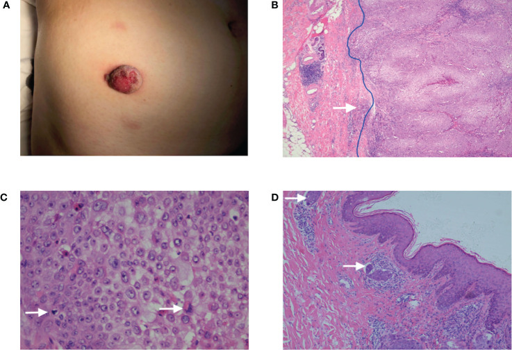
Figure 1.
(A) A reddish plaque on the skin of the right lower abdomen with a diameter of 2.5 cm. (B) The HE ×40: The tumor presented as an exo-endophytic proliferation with a dermal multinodular, well-circumscribed growth connected to the epidermis (blue line), and an invasive focus of frankly atypical epithelium could be observed (white arrow). (C) The HE ×400: The tumor was composed of clear, monomorphic cells with mitosis. (D) The HE ×100: Tumor thrombus could be observed (white arrow).

