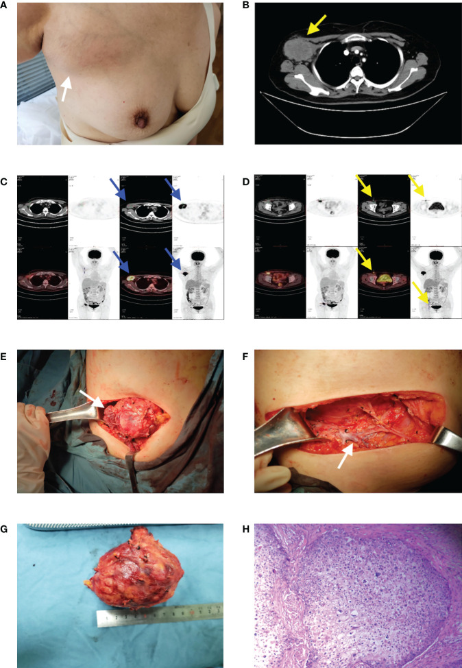
Figure 2.
(A) A mass in the right axilla. (B) Preoperative enhanced CT revealed a soft tissue density mass (yellow arrow) in the right axilla with a size of 6.9 × 5.2 cm. (C, D) PET-CT showed the mass as axillary lymph node metastasis (blue arrows) and another metastasis located at the ipsilateral inguinal lymph node (yellow arrows) which were characterized by shapes of nuclide accumulations in PET-CT image. (E) An intraoperative view from above, showing the tumoral invasion of the pectoralis major and minor muscles (white arrow). (F) An intraoperative view from above, showing the axillary vein (white arrow) after ALND. (G) The tumor was 7 cm in diameter. (H) The HE ×100 showed the tumor was characterized by a proliferation of tumoral lobules composed of large, atypical cells with clear cytoplasm.

