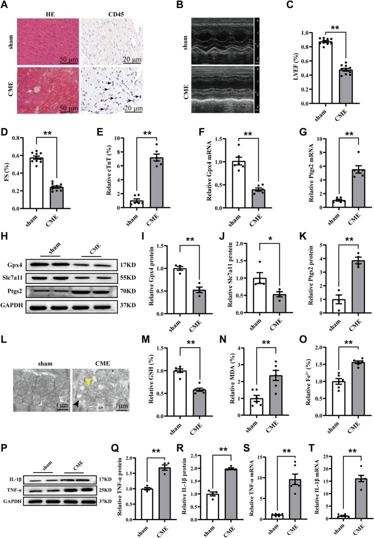
FIGURE 1.
Ferroptosis and inflammation involved in CME-induced myocardial injury. (A) Myocardial tissues stained with H&E (scale bar = 50 μm) and immunohistochemistry (scale bar = 20 μm). The white arrow indicates microspheres. The gray arrow indicates microinfarcts. The black arrow indicates the infiltration of inflammatory cells. (B–D) Cardiac LVEF and FS detected by echocardiography (n = 10). (E) Serum levels of cTnT in the sham and CME group (n = 6). (F,G) The expression levels of Gpx4 and Ptgs2 mRNA (n = 6). (H–K) Western blotting analysis of Gpx4, Slc7a11, and Ptgs2 (n = 4). (L) Representative images of transmission electron microscopy. The black arrow indicates the reduced or even disappeared mitochondrial cristae. The yellow arrow indicates the rupture of the outer membrane. Scale bar = 1 μm. (M–O) Cardiac levels of GSH, Fe2+, and MDA were measured (n = 6). (P–T) The expression of IL-1β and TNF-α in mRNA (n = 6) and protein levels (n = 4). GAPDH served as an internal control and was performed to quantitatively normalized the protein data. Data are presented as the normalized mean ± SEM (to sham) or mean ± SEM. Values in shams were averaged and normalized to 1 (E–T). *p < 0.05. **p < 0.01. CME: coronary microembolization; H&E: hematoxylin and eosin; LVEF: left ventricular ejection fraction; FS: fractional shortening; cTnT: cardiac troponin T; Gpx4: glutathione peroxidase 4; Slc7a11: solute carrier family 7 member 11; Ptgs2: prostaglandin-endoperoxide synthase-2; MDA: malondialdehyde; GSH: glutathione; IL-1β: interleukin 1 beta; TNF-α: tumor necrosis factor alpha.

