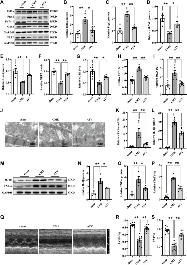
FIGURE 5.
Atorvastatin attenuated ferroptosis-dependent myocardial injury and inflammation following CME by inhibiting Hif1a/Ptgs2 axis. (A–F) Western blotting was performed to determine the protein levels of Hif1a, Ptgs2, Gpx4, Slc7a11, and NRF2 (n = 4). (G–I) Levels of GSH, Fe2+, and MDA in myocardial tissues (n = 6). (J) Representative images of transmission electron microscopy. Scale bar = 1 μm. (K,L) RT-qPCR was used to detect the mRNA expression of IL-1β and TNF-α (n = 6). (M–O) Western blotting showing the expression of IL-1β and TNF-α (n = 4). (P) Serum levels of cTnT were detected in each group (n = 6). (Q–S) Cardiac LVEF and FS detected by echocardiography (n = 10). GAPDH served as an internal control was performed to quantitatively normalized the protein data. Data are presented as the normalized mean ± SEM (to sham) or mean ± SEM. Values in shams were averaged and normalized to 1 (A–P). *p < 0.05. **p < 0.01. CME: coronary microembolization; Hif1a: hypoxia-inducible factor 1 subunit alpha; Ptgs2: prostaglandin-endoperoxide synthase-2; Gpx4: glutathione peroxidase 4; Slc7a11: solute carrier family 7 member 11; NRF2: nuclear factor erythroid 2-related factor 2; MDA: malondialdehyde; GSH: glutathione.

