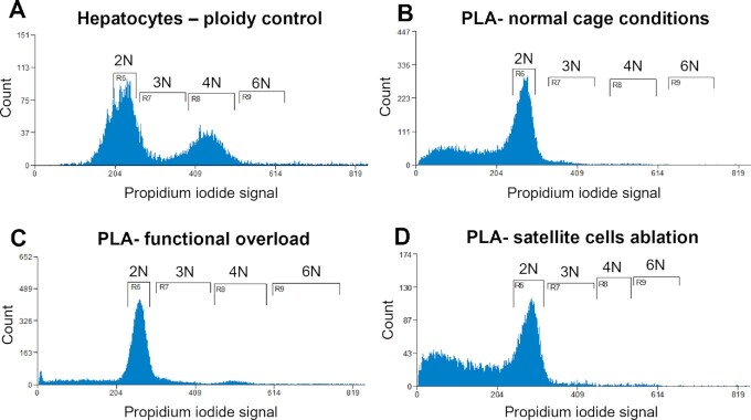
Figure 6.
Representative analysis of ploidy levels in sorted myonuclei from PLA muscles. Cell flow cytometry of hepatocytes (A), myonuclei isolated from PLA skeletal muscles from animals kept in the normal cage conditions (B), mice after functional overload (C), and mice after SCs ablation (D). The peaks corresponding to diploid nuclei are labeled 2 N, triploid nuclei as 3 N, tetraploid nuclei as 4 N, and hexaploid nuclei as 6 N.

