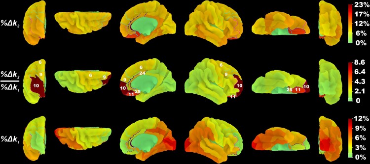
Figure 4.
Dissociation of k3 acetylcholinesterase hydrolysis versus k1 proxy flow changes in the anterior versus the posterior brain in the baseline Parkinson’s disease group. The percentage decrease in PMP k3 and k1 coefficients and the ratio of their percentage decrease from baseline visit to follow-up visit were mapped to BA volume-of-interests42 and projected to a high-resolution cortical surface mesh. Regions for which the visit model fitted k3 values significantly better than the confounding model are outlined and numbered based on their BA designation.

