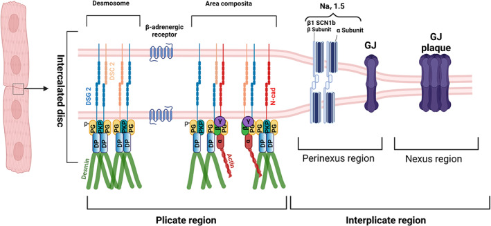
FIGURE 2.

Schematic of cardiomyocyte intercalated disc (ICD). The intercalated disc of the cardiomyocyte consists of plicate and interplicate regions, which in general lie perpendicular to each other. For convenience, we depicted here the two regions as parallel structures. The plicate region consists of the desmosome, as an individual mechanical component, and area composita or fascia adherens where both the desmosomes and adherens junction proteins are intermingled. Desmosomes are formed by the homo or heterophilic interactions of desmoglein 2 (DSG2) and desmocollin 2 (DSC2), where AJs are formed by the N‐cadherin (N‐cad). Desmosomes are anchored to the intermediate filament protein, desmin via the armadillo proteins plakoglobin (PG) also known as γ‐catenin and plakophillin (PKP) 2 which further interacts with the plaque protein desmoplakin, which then interacts with desmin. AJs are very well known to be anchored to the actin cytoskeleton via the alpha‐catenin (α), beta‐catenin (β) and the gamma‐catenin (known also as PG). But in the area composita, it could also be likely that desmosome and AJs are intermingled in such a way that desmosomes are anchored to the actin cytoskeleton and AJs to the desmin. In the interplicate region, there exists two regions, perinexus and nexus, respectively. In the perinexus region sodium channel complexes (Nav 1.5) concentrated at the edge of GJs, which are found in the nexus region, are involved in ephaptic conduction in the heart. The sodium channel β1 subunit with its extracellular adhesion domain plays a critical for this mechanism to function. Another important structure in ICD, that plays a major role in cardiomyocyte adhesion, is the β‐adrenergic receptor. Schematic was created with BioRender.Com
